
Figure 6.
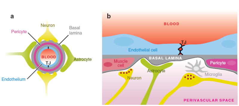
Schematic of the neurovascular unit. (a) A cross-section through a brain capillary shows adjacent endothelial cells connected by TJs that establish the BBB. The endothelial cell layer is surrounded by the basal lamina that separates the endothelium from the pericytes, astrocytes, and neurons. (b) A longitudinal section through a portion of a brain capillary reveals the presence of adjacent endothelial cells connected by TJs. Pericytes are present within the basal lamina in close proximity to the endothelial cells, whereas astrocytic endfeet are on the outer surface of the basal lamina. Microglia, nerve fibers, and neuromuscular synapses are found in the perivascular space. Panel b has been modified with permission from Abbott 2005; copyright 2005 by Springer Science and Business Media.