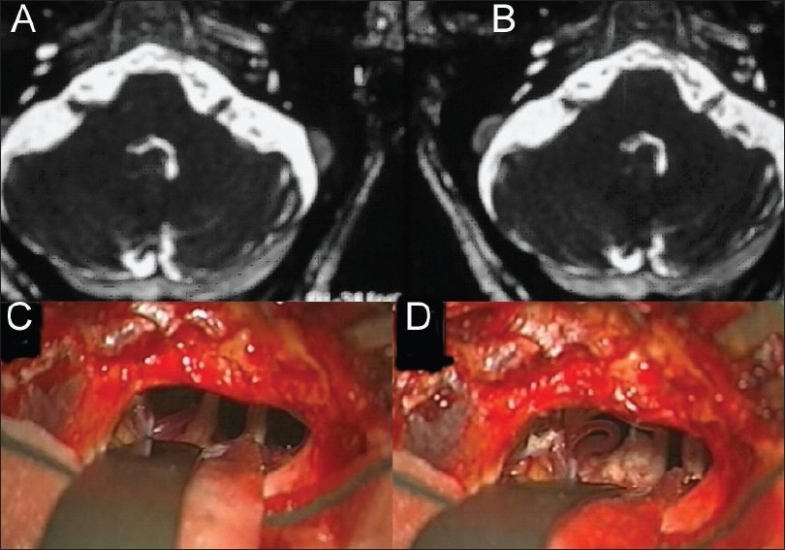
Figure 1.

67 yo male patient has a 7 years history of right trigeminal neuralgia and no response to regular medical treatment, above image is T2 weighted MRI that showed no significant changes Apart from high suspension of right vascular loop artifacts around trigeminal nerve entry zone. A and B pictures during surgery of the same patient: picture A there is a compression of the right trigeminal nerve by superior cerebellar artery, picture B showed separation of the artery and nerve with a piece of Teflon
