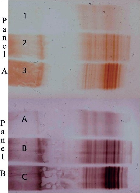
Figure 2.

CSF from nonMS (Lanes 1 and A) and 2 MS patients (Lanes 2, 3 and B, C) were examined by immunoblot analysis. Unconcentrated CSF was used in Panel A, and CSF diluted 1/10 used in Panel B. DAB enhancement with silver (Panel B) permitted dilution of CSF for analysis with excellent resolution of the OCB bands
