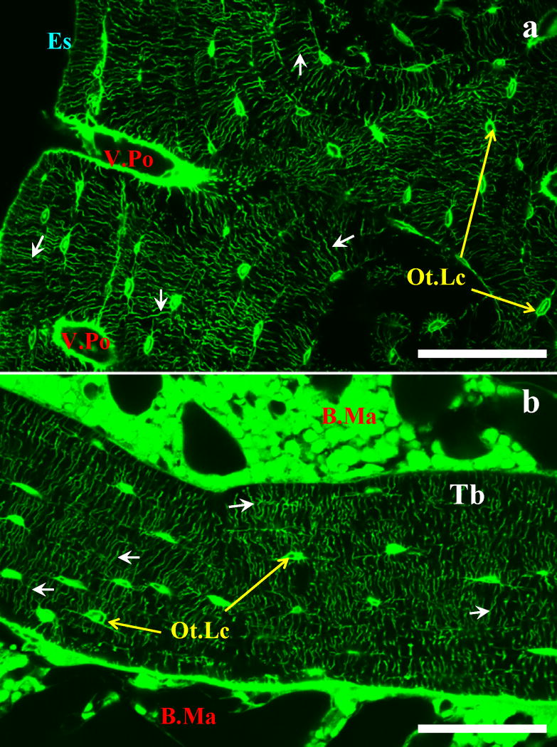
Figure 1.
(a) Unembedded cortical rat tibia diaphysis and (b) PMMA-embedded cancellous rat tibia metaphysis stained with FITC. The staining method demarcates the vascular porosity (V.Po), the canalicular porosity (small arrows), and the interstitial space surrounding the osteocyte lacunae (Ot.Lc) of both cortical and cancellous bone. Endosteal surface (Es), bone marrow (B.Ma), and trabecula (Tb) are also indicated; scale bars 130 μm.

