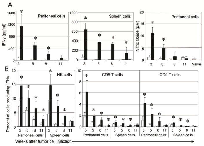
Figure 2. Tumor bearing mice treated with chNKG2D T cells have a sustained IFNγ response from host cells.
(A) ID8-GFP cells were injected i.p. and mice were treated with wtNKG2D T cells (white bars) or chNKG2D T cells (black bars) one week later. Three, five, eight, or eleven weeks after tumor cell injection, peritoneal wash cells and spleen cells from T cell treated or naive mice (grey bars) were cultured in media for 24 hours. Cell-free supernatants were collected and assayed for IFNγ or for nitric oxide. (B) Intracellular staining for IFNγ was performed on peritoneal wash cells and spleen cells cultured as described in (A). Cells were gated on either CD8+CD3+, CD4+CD3+, or NK1.1+CD3− as indicated. The average of each group (n=4) is shown + SD. Treatment with chNKG2D T cells significantly increased IFNγ and nitric oxide secretion compared to control treated mice (*-p<0.05). Data are representative of 2 separate experiments.

