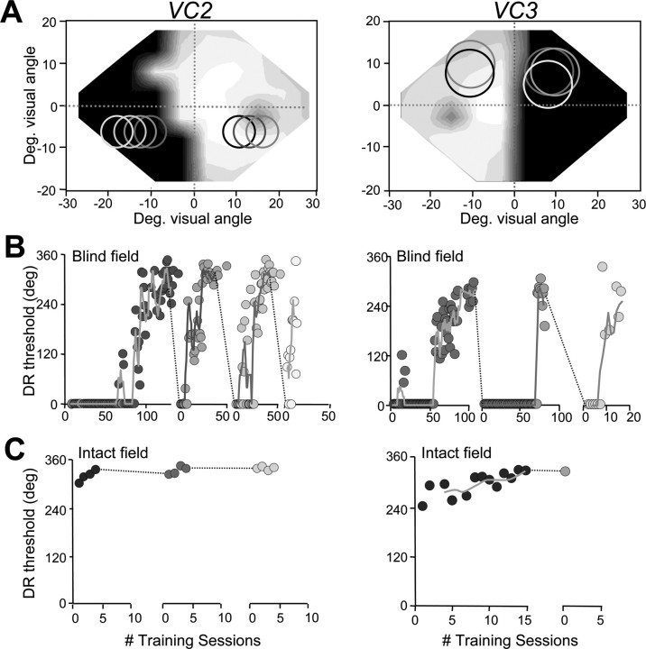
Figure 3.
Retinotopic specificity of training-induced improvements in direction range thresholds. A, Visual field maps for VC2 and VC3, illustrating the locations where visual training was performed. The shade of gray of circles in the visual fields match the shading of data points in B and C. B, Plots of direction range (DR) threshold versus the number (#) of training session at blind field locations in A. Once recovery of DR thresholds was attained at a given blind field location, moving the stimulus to a different location within the blind field, even one that was only 2° away, caused DR thresholds to fall to 0°. The process of retraining then had to be restarted anew. C, This contrasts with learning rates in the intact hemifields of the same subjects, in which DR thresholds ranged between 240 and 305° initially and stabilized to 330–340° with just a few days of training. However, this small improvement appeared to transfer very effectively when the stimulus was moved 2° deeper into the intact hemifield. Solid lines in the DR threshold graphs indicate moving averages with periods of four training sessions. Deg., Degree.

