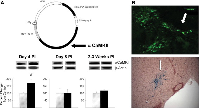
Figure 2.
Viral-mediated transient overexpression of αCaMKII in the NAcc shell. A, Western blots of αCaMKII obtained 4 d, 8 d, and 2–3 weeks postinfection (PI). αCaMKII was maximally and only elevated 4 d PI. Protein levels are expressed as group mean (+SEM) percentage change from controls (n = 3–6/group). *p < 0.05, αCaMKII (black bars) versus control (cross-hatched bars). The schema illustrates the components of the HSV amplicon used to overexpress αCaMKII. B, Photomicrographs of the NAcc obtained 4 d after infection with HSV-αCaMKII-GFP (top) or HSV-LacZ (bottom) illustrating GFP- or β-galactosidase-positive neurons in close proximity to the injection cannula tips (arrows).

