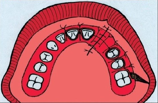
Figure 2D.

The bone graft covered with the palatal and gingival mucoperiosteal flaps. Note that the lateral gingival flap has been mobilised anteriorly to ensure good covering of the grafted area leaving the defect for secondary epithelialization in the region of the first molar.
