Figure 1.
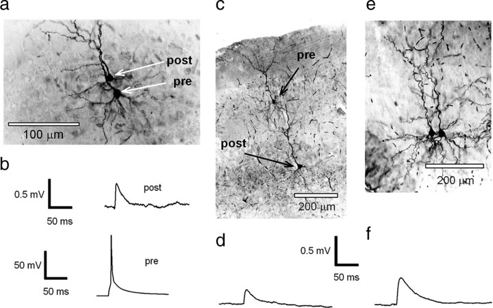
Recordings from synaptically connected pyramidal neurons in the cortex. a, Photomicrograph of a pair of L2/3 pyramidal neurons that were synaptically connected (see b). The neurons were filled with 0.5% biocytin during recording, and the tissue was subsequently processed by conventional methods to enable visualization of recorded neurons. b, Recordings made from the pair of neurons shown in a. Raw trace of a single action potential that was evoked in the presynaptic neuron by short pulse current injection (bottom) and the averaged postsynaptic EPSP recorded in the postsynaptic neuron (top). c, Photomicrograph of a layer 2/3 pyramidal neuron (presynaptic) that is synaptically connected to a layer 5 pyramidal neuron (postsynaptic), filled with 0.5% biocytin, visualized by conventional methods. d, Mean EPSP waveform recorded in a layer 5 neuron (c) evoked by an action potential generated in a presynaptic (L2/3) neuron. e, Photomicrograph of a pair of L5 pyramidal neurons that are synaptically connected, filled with 0.5% biocytin, and visualized by conventional methods. f, Mean EPSP waveform recorded in a postsynaptic L5 neuron evoked by an action potential generated in another L5 neuron.

