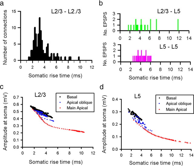
Figure 3.
Rise time as an indicator of dendritic location of EPSP. Distribution of experimental 10–90% EPSP rise times for recordings made from pairs of L2/3 pyramidal neurons (a), L2/3 to L5 pairs (b, top), and L5 to L5 connections (b, bottom) at room temperature. The effect of dendritic location on somatic EPSP amplitude and rise time was investigated using a representative L2/3 (c) and L5 (d) model neuron. Simulated EPSPs were generated using a brief injection of charge (0.1 pC) into every 50th spine across the entire dendritic arbor of the model neurons.

