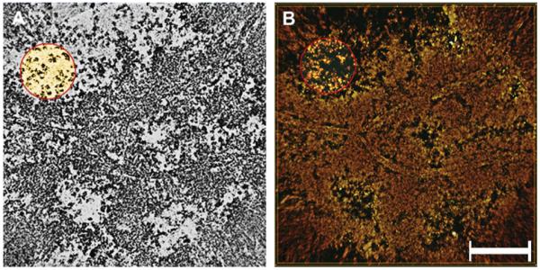
Fig. 1.

Reconstruction of cortical fibers from unextracted lenses. Panel A is a single-pixel slice (0.75 nm in thickness) cut along the X–Y plane showing the reconstructed field. The boundaries of three fibers are represented by plasma membranes. The cytoplasm contains a layer of electron-dense material associated with plasma membranes and less dense regions at the center, containing distinct particles arranged in clusters (circle). Panel B is the rendered map (~35 nm in thickness) depicting the overall distribution of plasma membranes and regions of greater and lesser density in the cytoplasm. Note that the particles seen in the single-pixel slices correspond to filaments (red circle). Bar: 0.2 μm.
