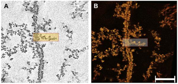
Fig. 4.

Cytoplasm after extraction of soluble proteins in a “ghost”. Panel A shows a single-pixel slice of the reconstructed field. The plasma membranes of fibers appear as trilayer structures representing the bilayer arrangement of phospholipids. The cytoplasm contains remnants of the matrix, which is composed of interlacing filaments distributed throughout. The filaments oriented themselves either parallel or perpendicular to the plane of the membrane. Individual filaments extend in zigzagging rather than straight paths and are studded with particles. Panel B shows a view of the rendered volume. Extraction of soluble proteins removed the clusters of particles seen in the denser regions in cortical fibers. The rectangles in panels A and B show a filament that is attached to the plasma membrane. Bar: 0.15 μm.
