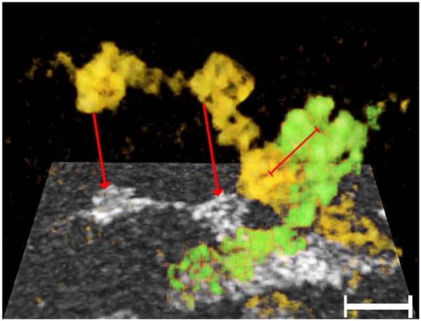
Fig. 6.

“Beaded” filaments forming the matrix in a “ghost”. The region where two filaments (yellow and green) cross is projected onto a single plane (rectangle below). The red arrows point to the projected representation of the corresponding rendered filament. Key structural properties of each filament are the thin core with pronounced kinks, and the cube-shaped particles studding its surface. At the region where the two filaments cross, particles from the respective filaments were in contact. The distance of the centers of these contiguous particles was ~12 nm center-to-center (red bar). Bar: 20 nm.
