Fig. 7.
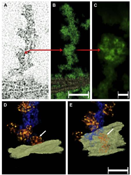
“Beaded” filaments and plasma membranes in a “ghost”. Panel A shows a slice of a “beaded filament” cluster that was oriented perpendicular to the plane of the plasma membrane. The red arrow indicates the location in the core where multiple filaments are separated by a small space. The red arrow also points to a particle decorating the filament on the left side. Panel B shows the rendered volume of the same bundle (green). The plasma membranes from neighboring fibers can be seen on the bottom. Bar: 50 nm. Panel C shows a higher magnification view of the particle indicated by the arrow in the single-pixel slice (panel A) and the rendered volume (panel B). Note that these particles contain an internal structure comprised of smaller globular domains. Bar: 6 nm. Panels D and E show a bundle of “beaded” filaments in order to demonstrate the association that the box-shaped particles (arrow) establish with the plasma membrane (white plane). Viewing the map at an angle from the extra-cellular space indicates that the particle associates with the plasma membrane at distinct points (arrows, panels D and E). Bar: 30 nm.

