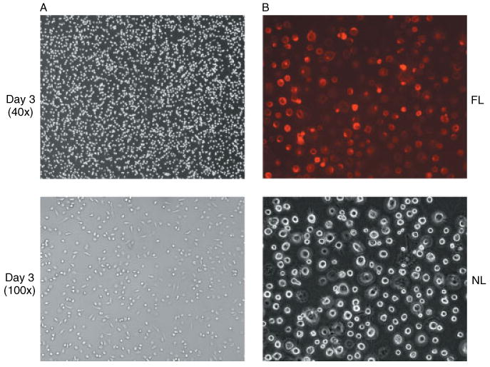
Figure 2.

Photomicrographs of primary cultures of human MDM. (A) Day 3 cultures of human MDM showing the formation of cell monolayers under phase-contrast microscopy. (B) Staining of primary cultures of human MDM at day 10 with mouse anti-human CD14 monoclonal antibody conjugated with R-phycoerytherin showing the same field under either normal light (NL) phase-contrast microscopy or fluorescent light (FL) microscopy (magnification 200×)
