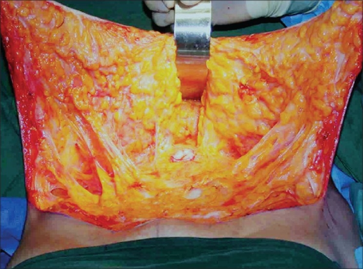
Figure 2b.

Inner surface of lipomobilized flap after selective undermining. Note the umbilicus in the centre and the retractor cranial to that. Midline plication is easily possible. Note the long strands of fibro-neuro-vascular connections and also the intact layer of adipose tissue left behind on the external oblique aponeurosis. This tissue is rich in lymphatics
