FIG. 1.
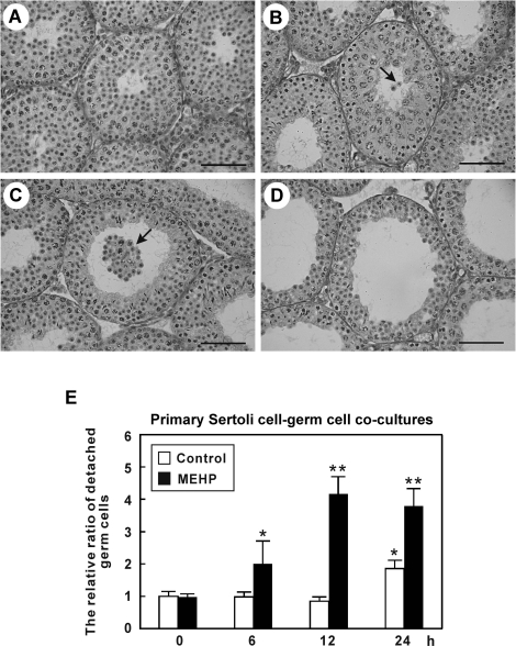
MEHP exposure causes germ cell detachment in vivo and in vitro. A–D) Testicular morphology in 28-day-old C57BL/6J male mice is shown in cross sections from paraffin-embedded testes with PAS-H staining. Detached germ cells are indicated by arrows. Control (A), MEHP 6 h (B), MEHP 12 h (C), and MEHP 24 h (D). Bar = 50 μm. E) Primary rat Sertoli cell-germ cell cocultures treated with or without 200 μM MEHP for 0, 6, 12, and 24 h, and detached cells were quantified. The open bar represents control cells and the solid bar represents MEHP-treated cells. Values represent the mean ± SEM. Asterisks denote significant differences between the treatments and the control (*P < 0.05, ** P < 0.01, Student t-test).

