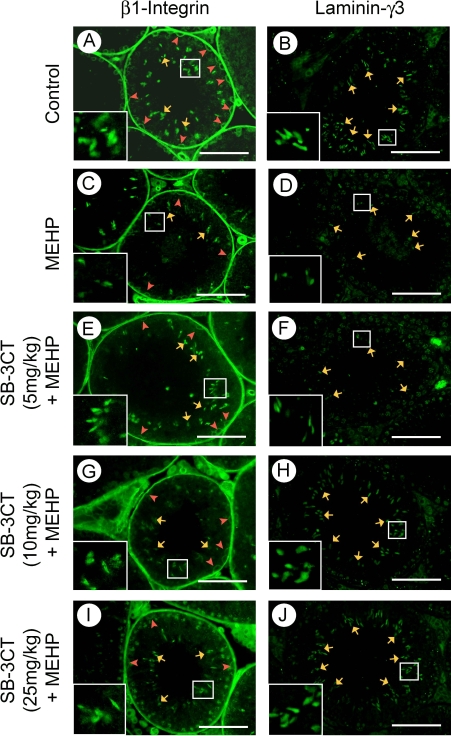
FIG. 7.
Dynamic changes in the expression and localization of β1-integrin and laminin-γ3 in the seminiferous epithelium are revealed by immunofluorescence analysis. Twenty-eight-day-old C57BL/6J mice are pretreated with SB-3CT (0, 5, 10, and 25 mg/kg) for 6 h and then posttreated with 1 g/kg of MEHP for another 12 h. Control (A and B); MEHP only (C and D); 5 mg/kg of SB-3CT pretreatment (E and F); 10 mg/kg of SB-3CT pretreatment (G and H); 25 mg/kg of SB-3CT pretreatment (I and J). Testis cross sections show that β1-integrin and laminin-γ3 express close to the acrosome of spermatids, where the ectoplasmic specialization is located (orange arrows). β1-integrin also strongly expresses along the basement membrane as well as close to spermatogonia (red arrow heads). The expression of both β1-integrin and laminin-γ3 is decreased after MEHP exposure while increased in SB-3CT-pretreated mice testes. Bar = 50 μm.

