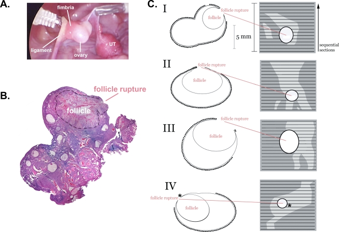
FIG. 3.
Schematic reconstruction of the extent of EpiX at the apex of the preovulatory follicle on four ovaries (I–IV) using a cytology brush (protocol 1). A) Photograph of ovary I during laparoscopic EpiX, showing the tip of the cytology brush, ovary, ovarian ligament, fimbria, and uterus (UT). B) Photomontage of a H&E-stained section from ovary I, showing the ruptured follicle (outlined with dashed line). C) Left diagrams represent schematics of representative sections from each ovary, containing the site of follicle rupture. The presence of OSE is denoted by (•). Right panels represent serial reconstructions showing the site of follicle rupture, intact OSE (dark gray), and brushed areas (light gray). Asterisk (*) indicates where a few OSE cells were present at the site of follicle rupture but were too few to be shown to scale in this schematic.

