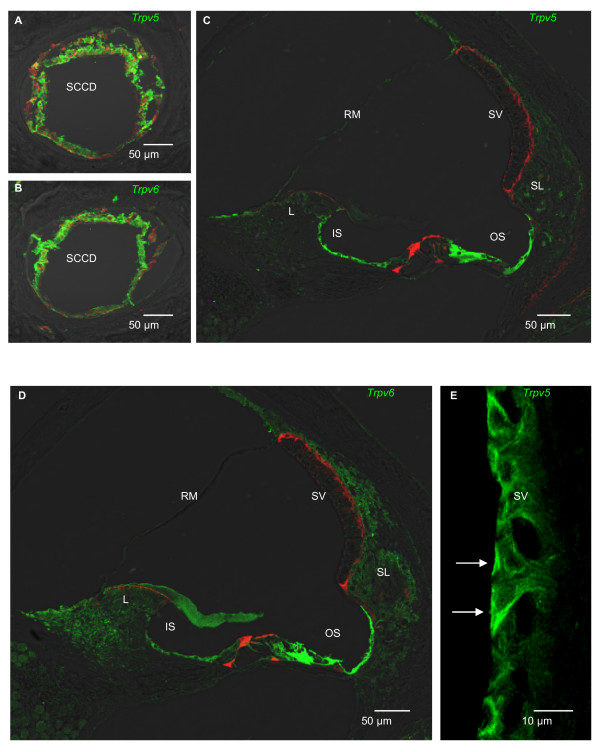
Figure 3.
Epithelial calcium channel immunolocalization in the cochlea and vestibular system of rat. Double-staining with an antibody against TRPV5 or TRPV6 (green) and actin (red; phalloidin) (A-D) and single-staining with an antibody against TRPV5 (green) (E). A, B) Cross sections of SCCD were stained against TRPV5 and TRPV6. C) Cross section of cochlea was stained against TRPV5. Outer sulcus cells (OS), inner sulcus cells (IS) and Hensen's cells (left of OS) show significant staining for TRPV5. D) Cross section of cochlea was stained against TRPV6. Outer sulcus cells, inner sulcus cells and Hensen's cells show significant staining for TRPV6. E) Cross section of stria vascularis (SV). Marginal cells of stria vascularis (arrows) show staining for TRPV5 at the apical membrane. Sections exposed to primary antibodies preabsorbed with antigenic peptide were not stained (not shown). RM, Reissner's membrane; L, spiral limbus; SL, spiral ligament.

