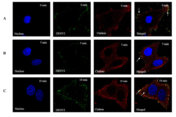
Figure 4.
Bio-imaging analysis of the interaction of clathrin molecules with DENV. DENV were stained green with anti-DENV E protein antibody conjugated to FITC, host clathrin stained red with anti-clathrin antibody conjugated to Texas Red (TR) and host nuclei stained blue with DAPI. (A) Attachment of DENV on the cell surface can be observed at 0 min p.i (arrow) with few co-localizations between DENV and clathrin molecules (arrows) (B) Obvious co-localization is observed between internalized DENV and clathrin by 5 minutes p.i (arrow). (C) Strong co-localization signals are observed between DENV2 and clathrin by 10 minutes p.i (arrows).

