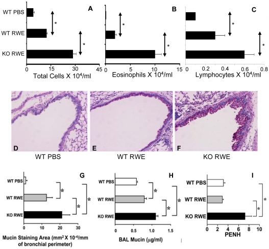
Figure 2. Role of FcγRIIb in allergic airway inflammation.
(A, B and C) Total inflammatory cells (A), eosinophils (B) and lymphocytes (C) were quantified in BAL of Balb/c RWE-sensitized WT and FcγRIIb KO mice challenged with either PBS (WT PBS) or RWE (WT RWE and KO RWE). (D, E and F) Lung sections were obtained from RWE-sensitized WT and FcγRIIb KO mice challenged with either PBS (WT PBS) or RWE (WT RWE & KO RWE). These sections were stained with PAS to identify mucin containing cells. (G) Mucin containing cells in the lung sections were analyzed by morphometric analyses of PAS staining area. (H) Mucin was quantified in BAL samples by ELISA using biotinylated mucin binding lectin. (I) WT and FcγRIIb KO mice were sensitized with RWE and challenged with either PBS (WT PBS) or RWE (WT & KO RWE). PENH was measured by Buxco whole body plethysmography. *, p<0.05.

