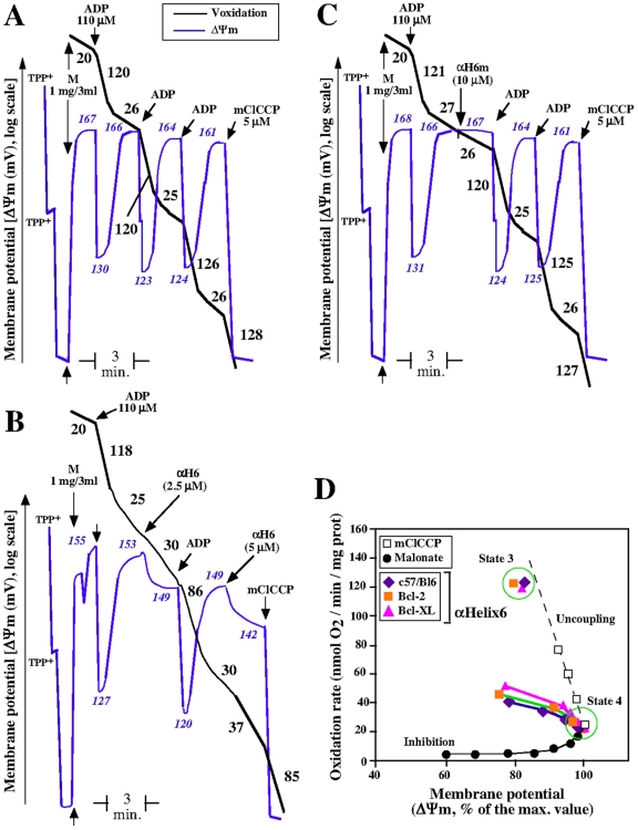
Figure 2. The helix αH6 affects mitochondrial bioenergetics.
Purified mice liver mitochondria (M) were incubated in respiratory buffer (0.33 mg/ml). Oxygen consumption (Voxidation, black line) and mitochondrial potential (ΔΨm, bleu line) were monitored using a Clark-type electrode coupled to a tetraphenylphosphonium (TPP+) cation-sensitive electrode, as described previously [13]. 10 mM of succinate was added as oxidizable substrate (malonate is 1 mM). ADP was added to 110 µM and mCCCP was added to 10 µM. The numbers along the traces gives the values of oxidation rates in nmol O2/min/mg protein (Black) and mitochondrial potential in mV (Blue) (A) Oxygen consumption and potential of mitochondria oxidizing succinate. (B) and (C) Effect of the addition αH6 and αH6m on the oxygen consumption and mitochondrial potential. Each panels (A), (B) and (C) is representative of 3 independent experiments. (D) Representation of the effect of αH6 on oxidation rate and potential of mitochondria isolated from control, Bcl-2 and Bcl-XL transgenic mice, as compared to uncoupling and inhibition of the mitochondrial respiration. Dotted black line shows the effect of the uncoupler CCCP, and the solid black line represents the inhibition of succinate-respiration by the specific inhibitor of complex II, malonate.

