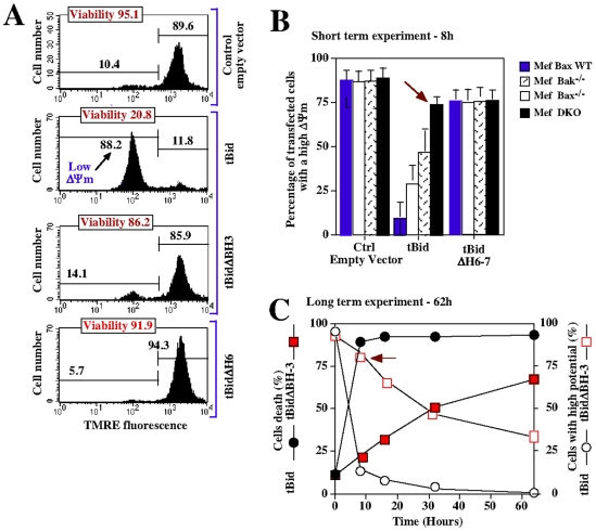
Figure 5. αH6 and BH3 domains are both required for tBid-induced apoptosis.
(A) Jurkat cells were transfected with either empty vector control or with plasmids encoding tBid, tBidΔBH3 and tBidΔH6. 12 h latter mitochondrial membrane potential ΔΨm (TMRE) and cell viability (PI) were assessed by FACS analysis. Each panels is representative of 3 independent experiments. (B) Wild-type, Bax+/+, Bax−/−, Bak−/− or double-knockout (DKO) Mefs cells were transfected with either empty vector control or with plasmids encoding tBid and tBidΔH6. After 8 h, green cells were analyzed by FACS for mitochondrial potential using TMRE. The red arrow indicates the ΔΨm in DKO Mefs. (C) Mefs cells were transfected with plasmids encoding tBid and tBidΔBH3, and the kinetics of mitochondrial depolarization and cell death were measured over 60 h by FACS analysis.

