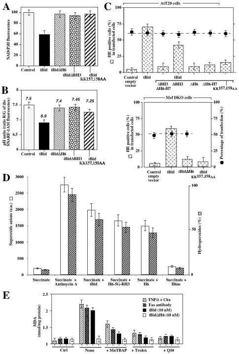
Figure 7. tBid required its helix αH6, but not its BH3 domain, to induce superoxide anion production and mitochondrial lipid peroxidation.
(A) and (B) Wild-type hepatocytes were transfected with a control vector (control) or plasmids encoding tBid, tBidΔH6, tBidΔBH3 and tBidKKAA and NAD(P)H and SNARF-1AM fluorescence (pH indicator) were measured by FACS. Data are given as % of the control ± SD. pH units were determined using a calibration curve generated using nigericin-permeabilized cells kept in buffer of different pH values. (C) AtT20 cells or DKO Mefs were transfected with empty vector (control) or plasmids encoding tBid, tBidΔH6, tBidΔBH3, tBid tBidKKAA, tBidΔH6–H7, tBidΔBH3ΔH6H7. Cells were then stained with hydroethidine (HE, Invitrogen/Molecular probes) to measure superoxide anion production. The percentages of transfected cells are indicated by the dotted line. (D) Purified mice liver mitochondria were energized using succinate (+ rotenone) and treated using 10 nM tBid, H6-5G-BH4, αH6 and αH6m. Mitochondrial superoxide anion production was measured using HE (a.u. = arbitrary units) whereas hydroperoxide was measured using amplex red. (E) Wild-type hepatocytes were treated with TNFα/cycloheximide, anti-Fas antibody or transfected with a plasmids encoding tBid and tBidΔH6. Mitochondrial lipid peroxidation was measured by FACS using MDA. We used the antioxidants trolox (2 mM), MnTBAP (1 mM) and MitoQ10 (1 µM).

