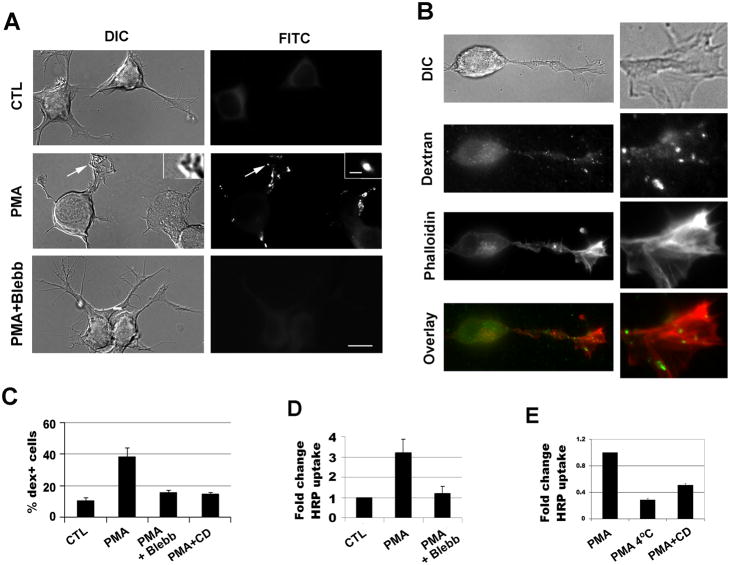
Figure 1.
PMA-induced macropinocytosis in Neuro-2a cells is inhibited by blebbistatin. (A) Neuro-2a cells were pre-treated with PMA for 3 minutes, followed by labeling with FITC-dextran for 2 minutes and the cells were subsequently rinsed and fixed. Labeling of dextran was examined by fluorescence microscopy and the localization in the reverse shadowcast vesicles was confirmed by DIC image. Preincubation with blebbistatin for 5 minutes before addition of PMA and FITC-dextran inhibited macropinocytosis. (B) Dextran uptake was carried out with PMA. Cells were washed and stained with phalloidin to visualize F-actin. Note that dex+ vesicles were localized in the neurites around the actin-rich membrane ruffles. 3X magnification of the images are shown to the right. (C) Percentages of dextran-positive cells were scored from >200 cells in each experiment and at least three independent experiments were carried out. (D, E) Horseradish peroxidase (HRP) uptake was also used to measure macropinocytosis with addition of vehicle control, PMA, or PMA with inhibitors. Blebb: blebbistatin; CD: cytochalasin D. Scale bar=10μm; scale bar in inset=1 μm.

