
Figure 5.
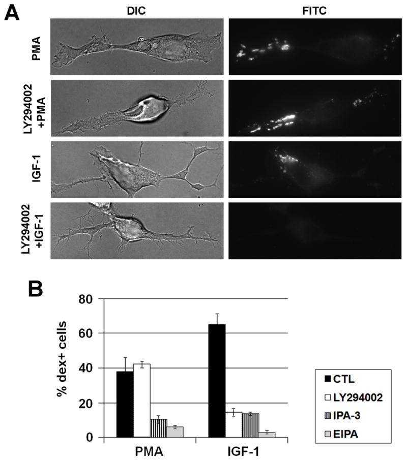
Comparison of macropocytosis induced by PMA and IGF-1 in Neuro-2a cells. A. Macropinocytosis was induced by PMA or IGF-1 as shown by 2 minute-dextran labeling. Note that the PMA-induced macropinosomes are located more in the neurites or processes whereas the IGF-1-induced macropinosomes are located in the membrane ruffles close to the cell body. The role of PI3K in macropinocytosis was analyzed by pretreatment with a PI3K inhibitor LY294002 for 30 minutes, prior to addition of IGF-1 or PMA. B. Percentages of dex+ cells induced by PMA or IGF-1 were quantified, in the presence of vehicle control, 50 μM LY294002, 10 μM PAK-1 inhibitor (IPA-3), or 100μM EIPA. Note that IPA-3 and EIPA inhibited both PMA and IGF-1-induced macropinocytosis whereas LY294002 inhibited only IGF-1-induced macropinocytosis.