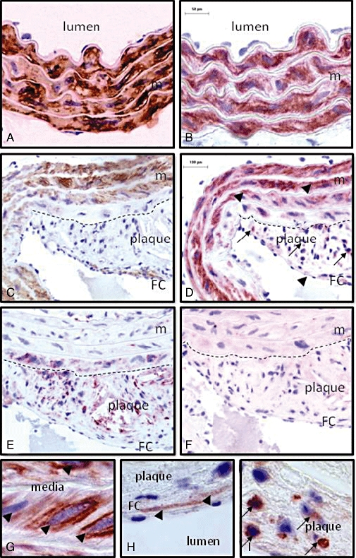
Figure 4.

Staining of α-smooth muscle actin (A, C, brown), of P2Y6 receptors (B, D, G, H, I, red) and of the macrophage marker mac-3 (E, red) in consecutive transverse sections of a plaque-free (A, B) and an atherosclerotic (C, D, E, F, G, H, I) segment of the aorta of an apoE–/– mouse (18 months old). α-Smooth muscle actin-positive staining was observed in the media of both segments, but also in SMCs of the fibrous cap. Mac-3-positive staining was found in the plaque, and occasionally in the inner media. P2Y6-positive staining was most abundant in SMCs of the media (arrow heads) in segments with (D, G) or without (B) plaques. However, in the plaque P2Y6 receptors were mostly present in macrophages (D, I, arrows) and in a few SMCs in the fibrous cap (D, H, arrow head). Staining of P2Y6 receptors was completely abolished after incubation of the antibody with the polypeptide that had been used to immunize the rabbits, illustrating the selectivity of the antibody (F). The dashed line shows the border between media and plaque. Magnification: 200× (A, B); 100× (C, D, E, F); 1000× (G, H, I). FC, fibrous cap; m, media; SMC, smooth muscle cell.
