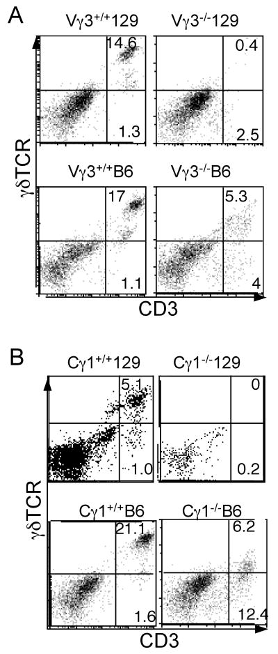
Figure 1.
Different developmental potentials of skin γδ T cells in Vγ3-/- or Cγ1-/- mice of 129 vs. B6 background. Epidermal cells were prepared from the skin of Vγ3 knockout (panel A) or 234JCγ1 (Cγ1) knockout mice (panel B) of 129 and B6 background and analyzed by flow cytometry for CD3 and γδTCR expression. The data shown in this figure, as well as in the other figures, are representatives of at least two independent experiments with similar results.

