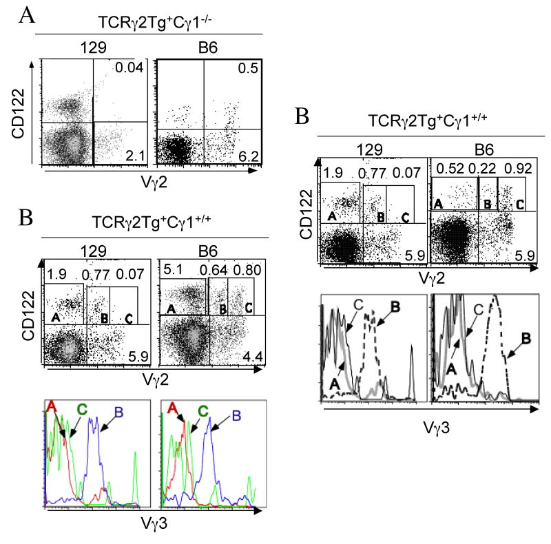
Figure 3.
Different positive selection processes of the transgenic Vγ2+ γδ T cells in fetal thymi of 129 vs. B6 background. A. Presence of positively selected transgenic Vγ2+ γδ T cells in the TCRγ2Tg+Cγ1-/- mice of the B6 but not 129 background. E16 fetal thymocytes of the TCRγ2Tg+Cγ1-/- mice of the 129 or B6 background were analyzed by flow cytometry for expression of CD122 on the transgenic Vγ2+ γδ T cells. B. The positively selected CD122+ Vγ2high+Vγ3- fetal thymic T cells were detected only in the TCRγ2Tg mice of the B6 background while the Vγ2medium+Vγ3+ fetal thymic T cells were positively selected in the TCRγ2Tg mice of both B6 and 129 backgrounds. E16 fetal thymocytes of the TCRγ2Tg mice of both backgrounds were analyzed by flow cytometry for expression of Vγ2, Vγ3 and CD122. The histographs in the top panels were gated on total thymocytes while the histographs in the bottom panels were gated on the CD122+ Vγ2− (A), CD122+Vγ2medium+ (B) or CD122+Vγ2high+ (C) population of the top panels (as indicated) and analyzed for Vγ3 TCR expression. Note that only the CD122+Vγ2medium+ population co-expressed Vγ3+ TCR. The CD122+ Vγ2- population (A) was mostly NK cells.

