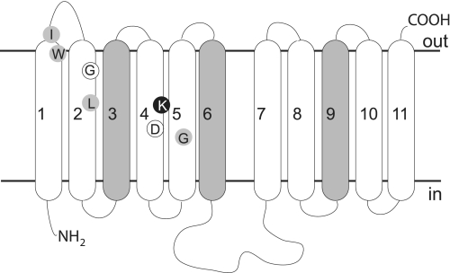
FIGURE 1.
Predicted topology of residues that influence ENT ligand specificity. The generic topology of an ENT family member is depicted, and N (NH2) and C (COOH) termini as well as intracellular (in) and extracellular (out) sides are indicated. Transmembrane helices predicted to lie on the exterior of the protein structure are shaded. Residues identified in this work are indicated in black (Lys155) and white (Gly86 and Asp159) circles. The predicted locations of residues described in the literature as influencing ligand specificity are indicated by gray circles (TM1, hENT2-I33 (51, 52) and hENT1-W29 (53); TM2, hENT1-L92 (54); TM4, LdNT1.1-K153 (15); TM5, LdNT1.1-G183 (55) and rENT1/rENT2 chimeras (56)).

