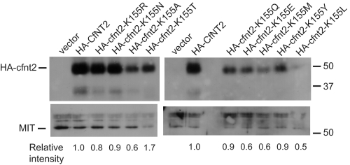
FIGURE 3.
Cell surface labeling of cfnt2-K155 mutants expressed in L. donovani. 4 × 10⁸ Δldnt1/Δldnt2 cells transfected with pALTneo carrying the indicated allele or with empty plasmid were treated with biotin as described under “Experimental Procedures.” Biotinylated proteins were bound to streptavidin beads and separated on a 10% SDS-polyacrylamide gel. Western blots were performed with anti-HA and anti-MIT antibodies, and proteins were visualized with chemiluminescence. Locations of size markers in kDa are indicated on the right, and the positions of HA-cfnt2 and the MIT protein (loading control) are indicated on the left. Relative intensities of the HA-cfnt2 bands were estimated from densitometry of scanned films.

