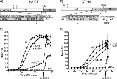
FIGURE 1.
Functional analysis of HA-CT and CT-HA. A and B, schematics of the N- and C-terminal peptide extensions are shown. Arg-192 (R192) is located at the cleavage site separating the A1- and A2-chains and bridged by a disulfide bond. Lys-4 and Lys-17 are indicated. * indicates the cleavage site for AcTev protease. C, time course of CT-induced Cl− secretion (Isc) induced by WT CT (filled square), HA-CT (filled triangle), or HA-CT pretreated with AcTev protease to remove the HA tag (filled circle). Some monolayers were pretreated with brefeldin A (indicated by + BFA) followed by application of WT CT (open square) or CT-HA pretreated with AcTev protease to remove the HA tag (open triangle). The viability of monolayers was demonstrated by applying the cAMP agonist, forskolin. D, exactly as for panel C but using the mutant toxin CT-HA. Error bars in C and D indicate S.D.

