FIGURE 6.
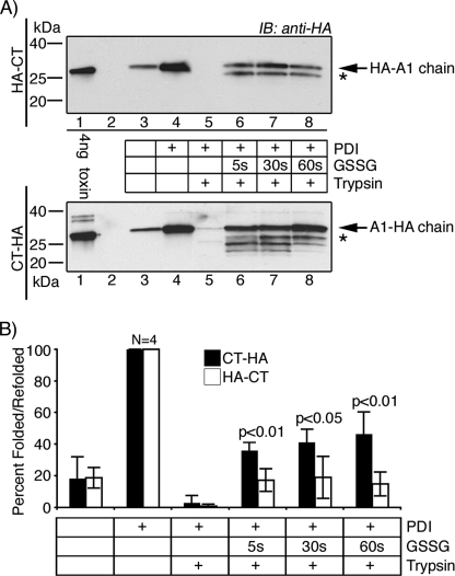
HA-CT refolds poorly after release from PDI in vitro. Briefly, we incubated all toxins with GM1-coupled beads and PDI in reducing conditions. PDI binds, unfolds, and releases the A1-chain into the supernatant from the B-subunit-GM1 magnetic beads. The supernatants were left untreated or were treated with GSSG to induce release of the A1-chain from PDI, and refolding was measured as resistance to trypsin degradation. All samples were analyzed by SDS-PAGE under non-reducing conditions, except the 4-ng control, which was reduced to separate the A1-chain. A, the unfolding and refolding of HA-CT (upper panel) and CT-HA (lower panel) were analyzed by immunoblot (IB) using an antibody against the HA tag. * indicates partially refolded toxin fragments. B, summarized data from four independent experiments. Compare HA-CT (white bars) with CT-HA (black bars). Band intensities were acquired using GeneSnap (GeneGnome HR, Syngene) and normalized to the total fraction of A1-chain released by PDI in the absence of trypsin (lanes 4). Error bars indicate S.D.

