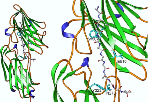
FIGURE 5.
Structural model of Fbl N2N3 subdomains in complex with γ-chain peptide. The fibrinogen-derived peptide is shown as a ball and stick object colored by atom type (carbon, gray; nitrogen, blue; oxygen, red). Residues Asn270, Tyr322, Trp507, and Glu510 are shown as stick objects in cyan. As in the case of ClfA, the conserved residues Tyr322 and Trp507 could help Fbl anchor the peptide through stacking interaction. Side chain atoms of Lys406 and Gln407 are not shown in the close-up view of the figure (right panel) for clarity. The backbone atoms of Asn270 and Glu510 of Fbl interact through hydrogen bonding interactions, which are shown as dotted lines.

