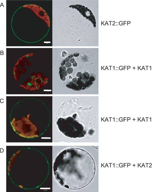
FIGURE 6.
Subcellular localization of KAT1-GFP in tobacco mesophyll protoplasts. A, protoplasts expressing KAT2-GFP alone (representative of 100% of GFP-stained protoplasts). B and C, protoplasts coexpressing KAT1-GFP and KAT1 (each representative of 50% of GFP-stained protoplasts). D, protoplasts coexpressing KAT1-GFP and KAT2 (representative of 90% of GFP-stained protoplasts). Left panels, protoplast sections analyzed for GFP fluorescence by confocal imaging; right panels, corresponding pictures obtained with transmitted light.

