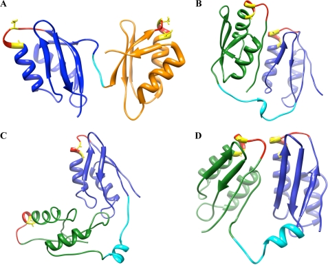
FIGURE 9.
Structures of pairs of neighboring MBDs. A, structure of MBD5 and MBD6 of ATP7B (adapted from Achila et al. (8); Protein Data Bank code 2ew9). B–D, ab initio structures for MBD1 and MBD2. MBD1 is shown in green, the inter-MBD loop in cyan, and MBD2 in blue. The GMxCxxC loop is shown in red, and copper-binding Cys residues are shown in yellow.

