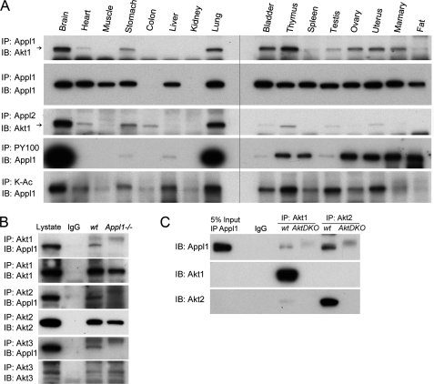
FIGURE 2.
A, Akt1 binds to Appl1 and Appl2 in a tissue-specific manner. Akt1 interacts with Appl1 in multiple tissues as demonstrated by using co-immunoprecipitation/immunoblot analysis. Akt1 and Appl2 also interact in various tissues. Co-IP also identifies tissue-specific phosphorylation and acetylation of the Appl1 protein, using phosphotyrosine (PY100)- and lysine acetylation (K-Ac)-specific antibodies. B, co-IP in wild-type and Appl1 knock-out MEFs. Akt1/2/3 were immunoprecipitated, and Western blot analysis was performed to detect Appl1. C, co-IP in Akt1/2 double knock-out (DKO) MEFs. Akt1 and Akt2 were immunoprecipitated, and immunoblotting (IB) was performed to detect Appl1.

