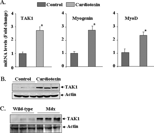
FIGURE 2.
Expression of TAK1 in regenerating TA muscle in vivo. A, TA muscle of 3-month-old C57BL6 mice was injected with saline alone or cardiotoxin as described under “Experimental Procedures.” After 5 days the TA muscle was isolated and processed for RNA isolation and measurement of mRNA levels for TAK1, myogenin, and MyoD by real-time-PCR. Data presented here show that the mRNA levels of TAK1 as well as myogenin and MyoD are significantly increased in cardiotoxin-injected regenerating TA muscle compared with contralateral saline-injected TA muscle (n = 3). *, p < 0.01, value significantly different from controls. B, representative immunoblots from two independent experiments presented here show that the protein levels of TAK1 are significantly increased in TA muscle 5 days after cardiotoxin injection. C, shown are protein levels of TAK1 in gastrocnemius muscle of 8-week-old wild-type and mdx mice measured by Western blot. The levels of TAK1 are noticeably higher in mdx mice compared with wild-type mice. There was no difference in the levels of unrelated protein actin.

