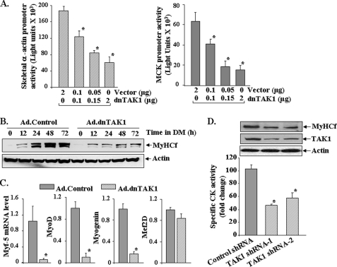
FIGURE 4.
Involvement of TAK1 in differentiation of C2C12 myoblasts. A, C2C12 myoblasts were transiently transfected with increasing amounts of dominant negative TAK1 (dnTAK1) plasmid along with either pSK-Luc or pMCK-Luc plasmid in a 1:10 ratio. After 24 h the cells were incubated in DM, and the luciferase activity in cell extracts was measured. Representative data from two independent experiments (each done in triplicate) presented here show that dnTAK1 inhibits the activation of both skeletal α actin and muscle creatine kinase promoters in a dose-dependent manner. *, p < 0.05, values significantly different from corresponding C2C12 cultures transfected with vector only. B, C2C12 myoblasts were transduced (multiplicity of infection 1:50) with control (Ad.Control) or dominant negative TAK1 (Ad.TAK1) adenoviral vectors for 24 h. The cells were then incubated in DM for indicated time intervals, and the expression of MyHCf was measured by Western blot. Representative immunoblots presented here show that dnTAK1 inhibits the expression of MyHCf without affecting the levels of an unrelated protein actin in C2C12 cultures. C, -fold difference is shown in the mRNA levels of Myf-5, MyoD, myogenin, and myocyte enhancer factor 2D (Mef2D) in Ad.control and Ad.dnTAK1-transduced C2C12 myoblasts 72 h after incubation in DM measured by real-time PCR technique. *, p < 0.01, values significantly different from C2C12 myoblasts transduced with Ad.control vector. D, C2C12 myoblasts were transfected with control or either of the two TAK1 shRNA plasmids, each containing a different target sequence for TAK1 knockdown. The cells were selected in the presence of puromycin (1.6 μg/ml) for 72–96 h followed by incubation in differentiation medium for 72h. CK activity in cell extracts was measured using the CK activity assay kit. The levels of MyHCf and TAK1 were measured by Western blot. Data presented here show that knockdown of TAK1 inhibits the expression of CK and MyHCf in C2C12 cultures. *, p < 0.01, values significantly different from control shRNA transfected C2C12 myoblasts.

