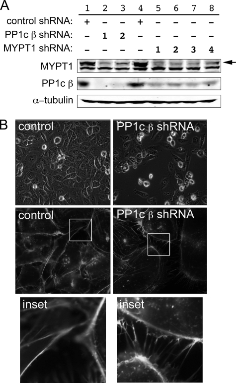
FIGURE 2.
Interdependence of PP1c β and MYPT1. A, CHO cells were infected with shRNAs directed against either PP1c β or MYPT1. 24 h following transfection, the cells were selected with blasticidin for an additional 2 days, rinsed with phosphate-buffered saline, lysed, and Western blotting was performed against the corresponding proteins using the indicated antibodies (see “Experimental Procedures”). Two independent shRNA sequences were used to knock down PP1c β and four for MYPT1, as indicated by the numbers in the legend. An immunoblot representative of two experiments with similar outcomes is shown. The top band seen in the MYPT1 Western blot (arrow) is MYPT1; the lower band is an unknown protein unspecifically immunostained by the primary antibody. B, morphological changes following PP1c β knockdown. MDA-MB-231 breast cancer cells were analyzed for changes in cellular morphology in the PP1c β knockdown cells using live cell imaging (top row) or using confocal microscopy with fixed cells stained with rhodamine-phalloidin to visualize F-actin (middle and bottom rows). 10 fields of cells were counted for each field for quantitation purposes. Figure is representative of two independent experiments.

