FIGURE 5.
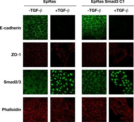
Stable expression of Smad3 does not inhibit TGF-β-induced EMT in EpRas cells. EpRas and EpRas S3 C1 cells were plated out at low density and either grown in the presence or absence of TGF-β1 (2 ng/ml) for a total of 10 days as described under “Experimental Procedures.” Cells were then processed for immunofluorescence using an anti-E-cadherin antibody, to analyze adherens junctions or an anti-Zona Occludens 1 (ZO-1) antibody, to analyze tight junctions. Smad2/3 localization and actin reorganization was visualized with an anti-Smad2/3 antibody and Texas Red-conjugated phalloidin, respectively. The E-cadherin and ZO-1 staining was performed on one sample of cells, and the Smad2/3 and phalloidin staining on another. In the EpRas S3 C1 cells, FLAG-Smad3 can be seen in the nucleus in the absence of TGF-β, as has been described for EGFP-Smad3 (56).

