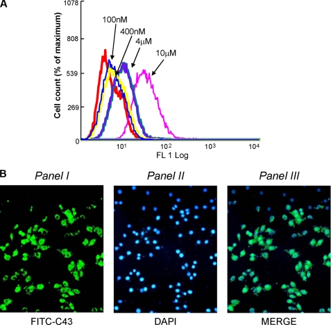
FIGURE 3.
Transduction of FITC-C43 into MCF-7 cells. A, MCF-7 cells were incubated with increasing concentrations of the fluorescein-conjugated peptide (FITC-C43), as indicated. Peptide uptake in MCF7 cells was detected by FACS analysis of fluorescein-labeled cells. The experiment was repeated four times with comparable results. The graph shows one representative experiment. FL, FITC log. B, MCF-7 cells were incubated with FITC-C43 peptide (10 μm) and fixed in formaldehyde at 37 °C. Uptake was monitored by fluorescence with appropriate filters. Panel I shows the FITC-C43 peptide internalized in the cells with prevalent submembrane localization; panel II shows cell nuclei stained with 4′,6-diamidino-2-phenylindole (DAPI) reagent; and panel III shows the overlay between the two images. These experiments were repeated four times with similar findings.

