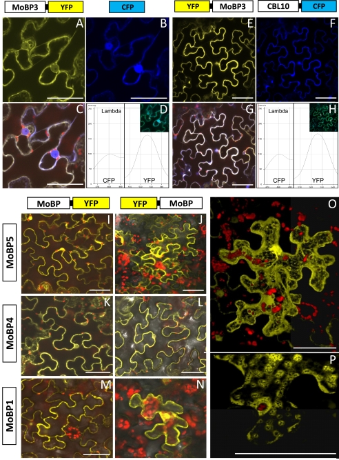
FIGURE 5.
Cytoplasmic localization of MoBP-YFP fusion proteins. cDNAs of MoBP1, -3, -4, and -5 were fused to the N and C terminus of YFP, respectively, and transferred via Agrobacterium infiltration into N. benthamiana leaves. A–H show co-localization with cytoplasmic marker proteins (CFP and CBL10::CFP); A and E, YFP fusion with MoBP3; B and F, CFP channel; C and G, merged pictures of A and B and E and F, respectively (the red color shows autofluorescence of the chloroplasts); D and H, spectral signature of CFP/YFP (peak at 480 and 525 nm, respectively) as detected in the λ mode. I–N show pictures of YFP fluorescence merged with the chlorophyll autofluorescence and the transmitted light photomultiplier in the channel mode of the confocal laser scanning microscope for MoBP1, -4, and -5 in both orientations to the fluorochrome. O and P, all scanning images of a single cell transformed with the MoBP3::YFP fusion construct are combined (Z-stack), the picture was taken at higher magnification and calculated using Volocity 4.4. Bars represent 50 μm.

