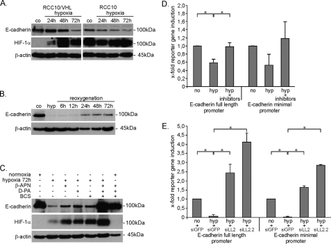
FIGURE 6.
Hypoxia leads to loss of E-cadherin expression, which requires LOX/LOXL2. A, E-cadherin expression was analyzed by immunoblotting in RCC10 cells after 72 h of hypoxia (1% O2). HIF-1α was analyzed in parallel to verify hypoxic induction. co, normoxic control. B, in RCC10/VHL cells E-cadherin expression was analyzed after reoxygenation, from 72 h of hypoxia. C, E-cadherin expression was analyzed in RCC10/VHL cells exposed to hypoxia for 72 h with or without treatment with pharmacological inhibitors of lysyl oxidases by immunoblotting. Hypoxia, 1% O2. For β-APN, d-penicillamine, and BCS, a 300 μm concentration of one was used or 100 μm each in combination. D and E, HKC-8 human renal tubular cells were transiently transfected with luciferase reporter constructs containing either the full-length E-cadherin promoter or a minimal promoter fragment containing two E-boxes of E-cadherin. D, 24 h after transfection, cells were stimulated with hypoxia (hyp), with or without the addition of lysyl oxidase inhibitors for a further 48 h (β-APN, d-penicillamine (d-PA), and BCS, each 100 μm). E, the day before reporter transfection, knockdown for LOXL2 with the two independent siRNAs siLOXL2 (siLL2) and siLOXL2.2 (siLL2.2) was performed. Controls were treated with siRNA against green fluorescent protein. Cells were exposed to hypoxia 24 h after reporter transfection. Results in D and E represent mean values of two independent experiments, with the error bars representing S.D. *, p < 0.05.

