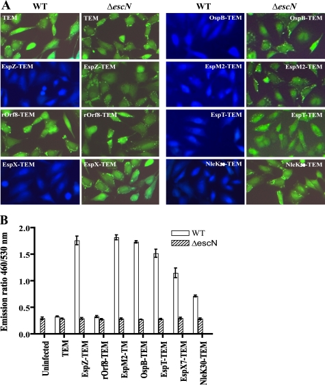
FIGURE 5.
Translocation of NleK, EspM2, EspT, OspB, and EspX7 into HeLa cells. A, HeLa cells were infected with EPEC WT and its ΔescN mutant carrying constructs expressing fusion proteins of EPEC EspZ and rOrf8, as well as C. rodentium EspX7, OspB, EspM2, EspT, and the N-terminal 30 amino acid residues of NleK, to β-lactamase TEM-1. The infected cells were loaded with CCF2/AM, and assessed for protein translocation using fluorescence microscopy with excitation at 409 nm. Blue fluorescence indicates positive type III translocation, and green fluorescence shows negative translocation. B, fluorescence quantification of HeLa cells infected by the same EPEC strains using a fluorescence plate reader. The data are averages with standard deviations of triplicate values of the results from one of two experiments, and are presented as the emission ratio between blue fluorescence (460 nm) and green fluorescence (530 nm).

