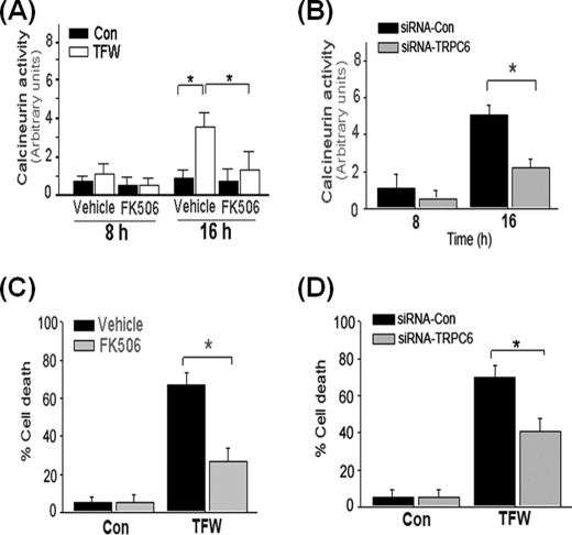
FIGURE 8.
TFW-induced cell death is dependent on TRPC6-mediated activation of calcineurin. A, time course of calcineurin activity in cultures of PC12 cells that were treated with FK506 (1 μm) 1 h prior to TFW. At the indicated time points following TFW, calcineurin activity in cell lysates was measured by a colorimetric assay using the RII phosphopeptide as substrate. Values are shown as arbitrary units and are the means ± S.E. of three independent experiments performed in duplicate. *, p < 0.05 (ANOVA with Scheffe post-hoc tests) compared with the indicated cultures. B, time course of calcineurin activity in cultures of PC12 cells that were treated with siRNA-Con and siRNA-TRPC6 (100 nm) for 18 h prior to TFW. The values are the means ± S.E. of three independent experiments performed in duplicate. *, p < 0.05 (ANOVA with Scheffe post-hoc tests) compared with the indicated cultures. C, cultures of PC12 cells were exposed to the calcineurin inhibitor FK506 (1 μm) 1 h prior to TFW. Cell viability was determined 24 h after TFW by Hoechst staining. Forty fields compromising at least 30–50 cells were counted. Results shown are the means ± S.E. of three independent experiments performed in duplicate. *, p < 0.05 (ANOVA with Scheffe post-hoc tests) compared with vehicle-treated cultures. D, cultures of PC12 cells were exposed to siRNA-Con and siRNA-TRPC6 (100 nm) for 18 h prior to TFW. Cell viability was determined 24 h after TFW by Hoechst staining. The values are the means ± S.E. of three independent experiments performed in duplicate. *, p < 0.01; **, p < 0.05 (ANOVA with Scheffe post-hoc tests) compared with the indicated cultures.

