
Figure 2. Proliferation of Tg and Tgmut B cells in response to anti-CD40 and anti-IgM stimulation.
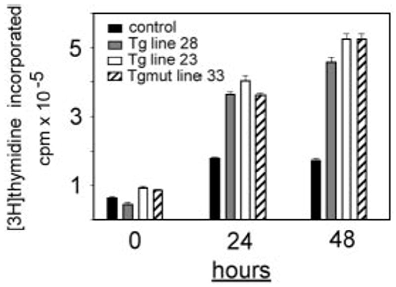
B cells from spleens of littermate control (■), Tg line 28 ( ), Tg line 23 (□), or Tgmut line 33 (▨) or were stimulated in vitro for 24 or 48 hours with anti-CD40 and anti-IgM. Incorporation of [3H] thymidine is shown with standard deviation (n = 4) for each bar.