
Figure 4. Tg-driven splenic lymphoma and peripheral leukemia.
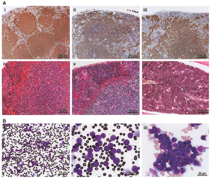
(A) A B220-stained section of asymptomatic Tg spleen (i; bar, 200 μm) is compared with a B220-stained section of a Tg mouse (ii; bar, 100 μm) or Tgmut mouse (iii; bar, 100 μm) with lymphocytic lymphoma. An H&E-stained section of asymptomatic Tg spleen (iv; bar, 50 μm) is compared with an H&E-stained section of a Tg mouse with lymphocytic lymphoma (v; bar, 50 μm). An H&E-stained section of bone marrow of Tgmut mouse with lymphocytic lymphoma is also shown (vi; bar, 100 μm). (B) A Wright-Giemsa–stained smear of peripheral blood of a leukemic mouse that received Tg B-cell lymphoma transplants shows overproliferation of lymphoblasts (i; bar, 50 μm). Blasts exhibit prominent, multiple chromocenters and scanty dark blue cytoplasm (ii; bar, 20 μm). Note the polymorphonuclear granulocyte for comparison. Additional field emphasizes homotypic adhesion and mixed chromatin states (iii; bar, 20 μm).