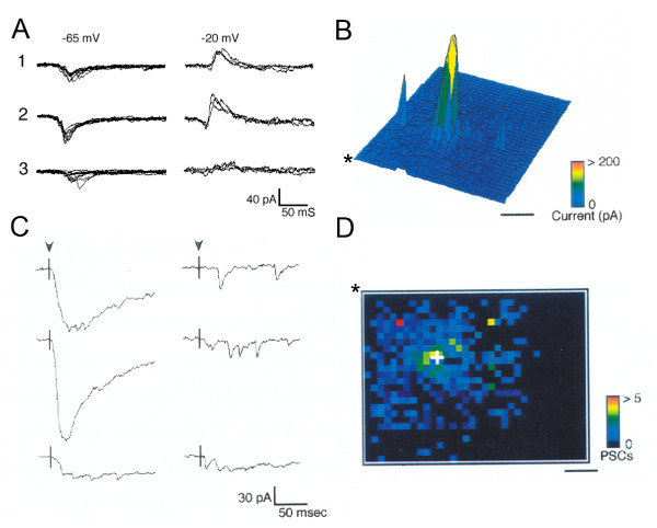
Figure 1.
Methods for visualizing synaptic inputs using photostimulation. (A) Inhibitory and excitatory inputs can be distinguished by shifting the holding potential of the recorded neuron. Traces from photostimulation of three different sites shown with holding potential set at -65 mv (left) or -20 mv (right). Each site is stimulated several times at each holding potential to illustrate the stereotyped response to repeated photostimulation. Site one is scored as inhibitory only. Site two is scored as excitatory and inhibitory. Site three is scored as excitatory only. (B) Amplitude plot of a map of 982 stimulation sites covering an area of 2.0 mm2 in a tangential slice of young (postnatal day 30) ferret visual cortex. The amplitude of the response at each site is coded by the color and height of the peak. The large central peak is due to direct activation of glutamate receptors on the recorded neuron. (C) Traces from stimulation of a few sites in the map shown in (B). On the left are traces from near to the recorded neuron containing direct and synaptic currents. Evoked synaptic currents are typically of similar amplitude but can vary in number. On the right are traces from stimulation of sites distant from the recorded neuron. Many stimulated locations do not generate a direct or synaptic response. (D) Plot of the number of synaptic currents evoked by stimulating each site. The post-synaptic current (PSC) plot shown is the result of analyzing the map in (B). In (D) the map is rotated and the three-dimensional perspective removed relative to (B). The asterisk is located at the same corner of the maps in (B) and (D). This plot enables the patterns and locations of inputs to be easily discerned. Scale bars = 500 μm.

